
Understanding the microscopic world of cells and bacteria is very important, but it is difficult to study in detail without harming the subject. EPFL researchers have developed a new microscope technology that combines two existing microscope technologies. This allows scientists to create HD and 3D images inside and outside the cell.
There are many different microscopic imaging techniques available to scientists, but they all have their strengths and weaknesses. An electron microscope can reveal complex details about the surface of a specimen, but it cannot be used for living cells because the intensity of the electron beam destroys the sample. Other methods, such as fluorescence microscopy, do not harm the sample, but separation is costly.
Therefore, for new research, EPFL researchers began by developing their own imaging technology. It is based on an existing one called scanning probe microscopy, in which the sample is jabbed with a probe tip to map the surface. However, because it invades cells, the EPFL team replaced the probe with a glass nanopore that measures ion flow without touching the sample. They called this method the Scanning Ion Conductance Microscope (SICM).
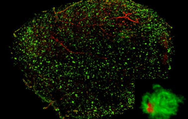
The team combined this new SICM technology with an existing technology called Stochastic Optical Fluctuation Imaging (SOFI). It allows you to look inside the cell and monitor the various molecules and processes that are going on there. The combination of the two technologies allows scientists to simultaneously capture high-resolution 3D images of the inside and outside of cells.
The team’s new microscopic imaging technology allows scientists to observe a variety of cellular processes
LBNI-LBEN
“The cell membrane is where it interacts with its surroundings,” says study author Samuel Mendes Reitan. “Here, as in the case of cell infection, many biological processes and morphological changes occur. With our system, researchers analyze intracellular molecular sequences and they are associated with membrane dynamics. You can map how they correlate. “
Perhaps most importantly, you can monitor the process over time, on a scale of less than a second to a few days. In the test, the team was able to observe mammalian cells move around, communicate, differentiate, swallow molecules through membranes, and infect bacteria.
Researchers say the new technology will be a very useful tool for infectious biology, immunology, neurobiology, but also in other areas such as energy science to support solar fuel production and more. It states that it can be used.
Leave a Reply